WELCOME TO CREATIVE BIOARRAY
WELCOME TO CREATIVE BIOARRAY
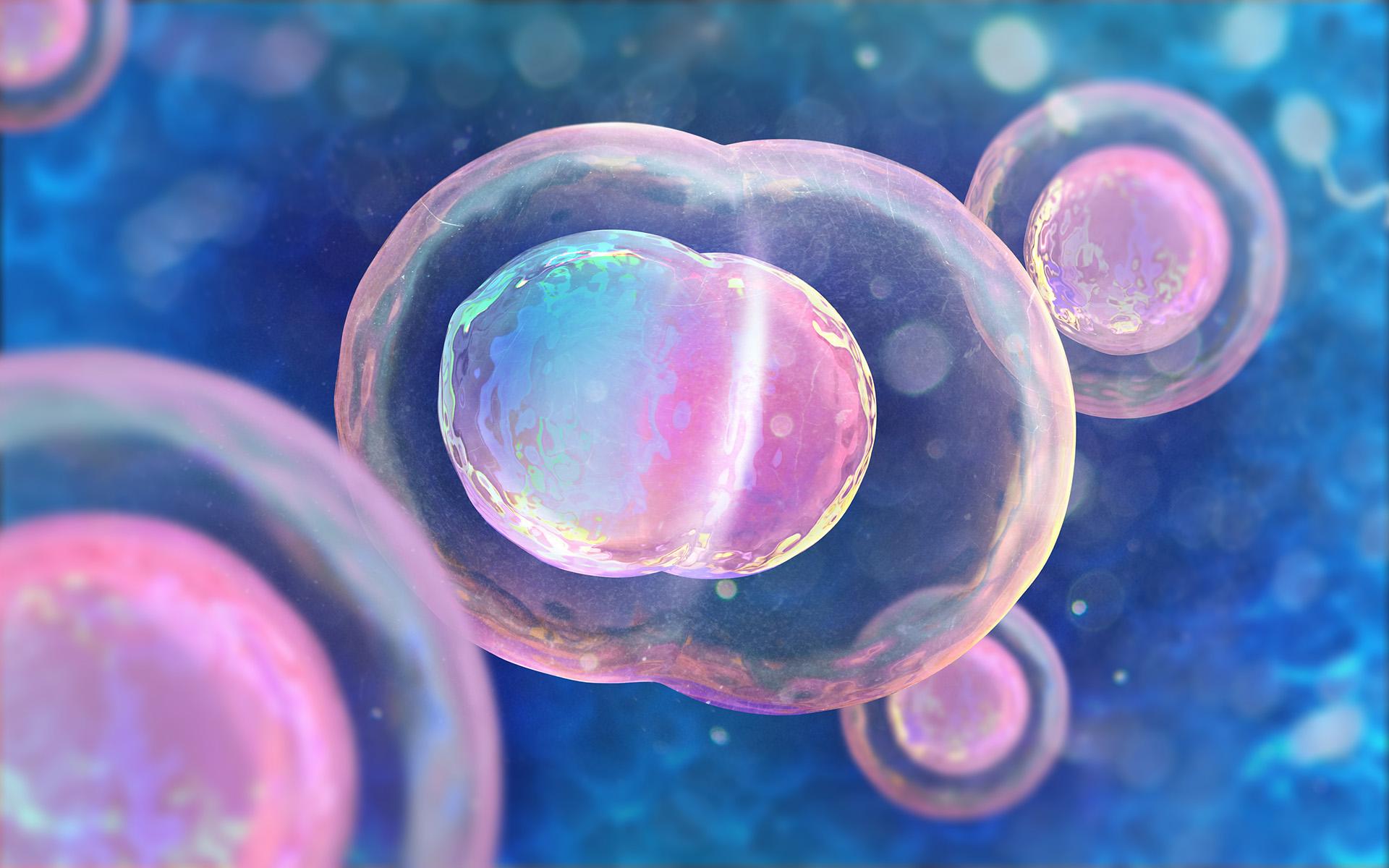
As cell research advances, so does the need for cell imaging technology. Today's researchers need a continuous flow of dynamic, long-term experiments on cell migration, interactions, and responses to environmental disturbances that cannot be met by conventional microscopes that can only observe stationary cells. Creative Bioarray' Olympus FV3000 Confocal Microscope-based live cell imaging technology can meet your stringent experimental needs.
The Olympus FV3000 Laser Scanning Microscope is a fully spectral microscope with a brand new TruSpectral Technology that combines high performance imaging capabilities with customisable software that is easy to use. Main features include: Fast and accurate imaging using a high-speed scanners, bright fluorescent imaging of even dim samples due to high-sensitivity detectors and improved observation reliability achieved by precise spectroscopy and newly developed time management system.
1. The FV3000 enables macro scale imaging from 1.25× while retaining capabilities of super resolution imaging at up to 150× magnification. 2. Silicon immersion objectives for accurate 3D imaging. 3. Resonant scanner to capture highly dynamic biological processes. 4. Multi-Dimensional Time-lapse with Z-drift compensator. 5. 3D Mosaic Imaging
The FLUOVIEW FV3000 series is designed to meet some of the most difficult challenges in modern science. Featuring the high sensitivity and speed required for live cell and tissue imaging, the FV3000 enables 2D-6D (x,y,z,t,λ,p) macro-to-micro imaging of cells, tissues, and small organisms. With an intuitive and adaptable user interface, the FV3000 supports complete workflows from image acquisition to processing and analysis. Particular attention has been paid to the needs of cell biology, cancer research, and stem cell research, and with two new upright configurations, the FV3000 is also poised to meet the needs of neuroscience, electrophysiology, and developmental biology.
1. Fluorescence spectral unmixing (up to 16 channels) 1) 2D data histogram display 2) Ratio imaging and IMD display 3) Fluorescence intensity and time-lapse measurements 4) Rolling average processing 5) Projections (including maximum intensity Z) 6) Z gap correction 7) Image editing (including individual color setting, pseudo-color, and graphic/text annotation) 8) FRET 9) FRAP 10) Super-Resolution 11) Noise Reduction 2. Featured cellSens Image Processing and Analysis Functions 1) Constrained Iterative Deconvolution 2) Count and Measure (Object Segmentation, Measurement, Counting, Classification) 3) Kymography (Velocity Measurements) 4) One-click macros for automated processing and analysis
The Olympus FV3000 Confocal Microscope-based cell imaging service offered by Creative Bioarray supports high-resolution fluorescence and brightfield image acquisition, enabling real-time recording of data over hours, days, or weeks. If you need our help, please contact us directly.
For research use only, not intended for any clinical use.
Please fill out the form below and we will get back to you as soon as possible with a quotation for the item you are interested in.